Before Your Operation
Before your operation your doctor or nurse will take a detailed medical history, including any medications you are currently taking. Please prepare your list of medications. You may also need to undergo tests such as blood work and an ECG to ensure you are medically fit for the procedure.
If cataract removal is planned as part of your surgery, additional measurements will be taken to determine the correct lens implant needed for successful visual outcomes.
At this appointment, we will also explain the surgical procedure in detail and answer any questions you may have. Additionally, we will review your medications and provide guidance on whether any need to be stopped prior to surgery, particularly blood thinners and medications used to manage diabetes.
What to Expect on the Day of Surgery
You will receive specific instructions about when to stop eating and drinking before your operation. Depending on the scheduled time of your surgery, you will be asked to arrive either early in the morning (for morning procedures) or later in the morning (for afternoon procedures).
In most cases, an overnight stay in the hospital is required, so we recommend bringing a small overnight bag with toiletries and a change of clothes.
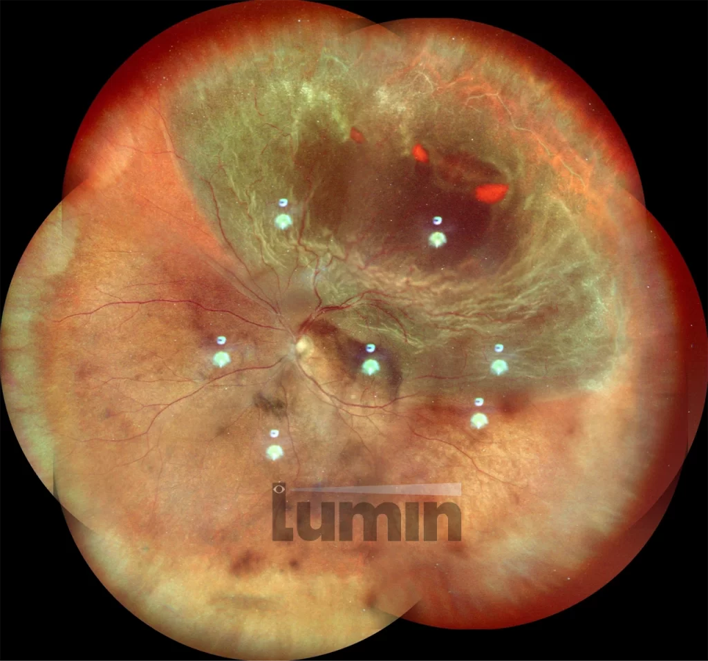
Please be aware that retinal detachment surgery may sometimes need to be performed as an emergency, depending on the urgency of your condition.